Posted by: Atlantic Eye Institute in Education

There are three types of bumps you may notice in or around the eye.
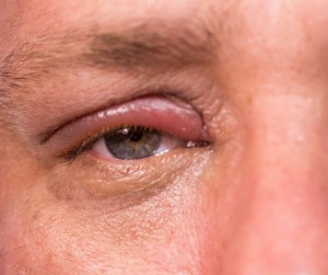
1. A stye (or hordeolum) appears as a pimple on the margin of the eyelid around the root of an eyelash. It is an infection that originates in the oil gland of an eyelash. This can be caused by make-up, dust, or debris getting into the gland and clogging it.
Typically a stye can be associated with redness, tenderness, pain, as well as, discomfort when blinking the eyelid. If infected, a bit of pus (appearing as a small, yellowish discharge) may also be present, and if the infection spreads beyond the gland, the skin and/or eye may be red.
In most cases, a warm compress can soften the stye’s contents and let it drain more easily. Run warm water over a clean washcloth. Wring out the washcloth and place it over your closed eye. Re-wet the washcloth when it loses heat; continue this for 5-10 minutes. Then, gently massage the eyelid. Doing this 2-3 times a day may help the stye to drain on its own. Sometimes antibiotic preparations are needed, and in rare cases an incision and drainage is necessary. Recurrence is likely if chronic underlying conditions are not addressed such as blepharitis, a chronic inflammation along the edge of the eyelid or rosacea. Your doctor at Atlantic Eye Institute may recommend daily cleansing of the eyelids and eyelashes with a gentle soap (such as baby shampoo) or Avenova eyelid cleanser.
If you have a stye it is important to avoid squeezing and poking the stye, because it could lead to scarring of the eyelid or spread the infection. Do not pluck your eyelashes to get rid of the stye, this can cause other problems. Gently wash the affected eyelid with mild soap and water. It is a good idea to avoid wearing eye makeup until the stye has healed. Contact lenses can be contaminated with bacteria associated with a stye, try to go without them until your stye goes away.
To reduce your risk of getting a stye be sure to wash your hands before touching your eye. Thoroughly disinfect your contact lenses. Always take your make-up off at night. Replace your cosmetic products every 6 months.
2. A chalazion appears as a swollen area or a small lump on the upper or lower lid. They are caused by an obstruction in a gland that is located in the eyelid tissue. This gland produces the lubricating tear film that keeps the eye moist. The obstruction results in retention of tear fluid inside the gland and swelling of the lid. Usually chalazions are painless, but feel tender during the early stages. Occasionally they grow to a size of a small fingernail and become rigid. In rare cases the chalazion may grow large enough to apply pressure on the eye and cause blurring of vision.
A chalazion is not an infection but it can become the sight for an infection if left untreated. The exact cause is unknown, but they tend to be associated with dry flaky skin, dry eyes, chronic inflammation and acne.
Typically chalazia will disappear without any treatment. Hot packs and drops may be helpful during the early stages. In rare chronic cases it may be removed, drained, with a simple in office procedure.
3. Pterygia and pingueculae are abnormal growths on the surface of the eye. A pterygium is a fleshy wedge shaped growth on the cornea, clear front part of the eye. This is a result of an abnormal tissue growth that usually begins on the inner corner of the eye. Typically, pterygium results from not having adequate UV protection; it is incredibly important to always wear UV protective sunglasses when you are outside. Pterygium can also be caused by chronic dry eye, or exposure to wind and dust.
Symptoms of pterygium are typically not severe but may include blurred vision, itching, burning and scratchiness.
Treatment is not necessary if there are no noticeable symptoms. If the pterygium becomes red or irritated, eye drops or ointments are used to reduce inflammation and relieve dryness. If good vision is threatened, a pterygium can be surgically removed. Pterygia have a tendency to return especially in younger people. In addition, the symptoms of dryness and irritation often persist after removal. Surface radiation or medications can be used to help prevent recurrences.
A pinguecula appears as a yellowish or white lump in the sclera or white part of the eye. It is composed of benign material, such as fat or degenerated tissue. This differs from the pterygia because it never grows over the cornea and is separated from the cornea by normal tissue. The pinguecula usually does not interfere with sight. Pingueculae are usually caused by dryness and exposure to the environment. Usually no symptoms occur, but you may experience some stinging and burning and your eye may become red or irritated by smoke, dust or wind. If it becomes inflamed drops are used to clear redness and irritation. They are rarely required to be surgically removed, but can be for cosmetic purpose. The down side it that pinguecula frequently returns after removal.


