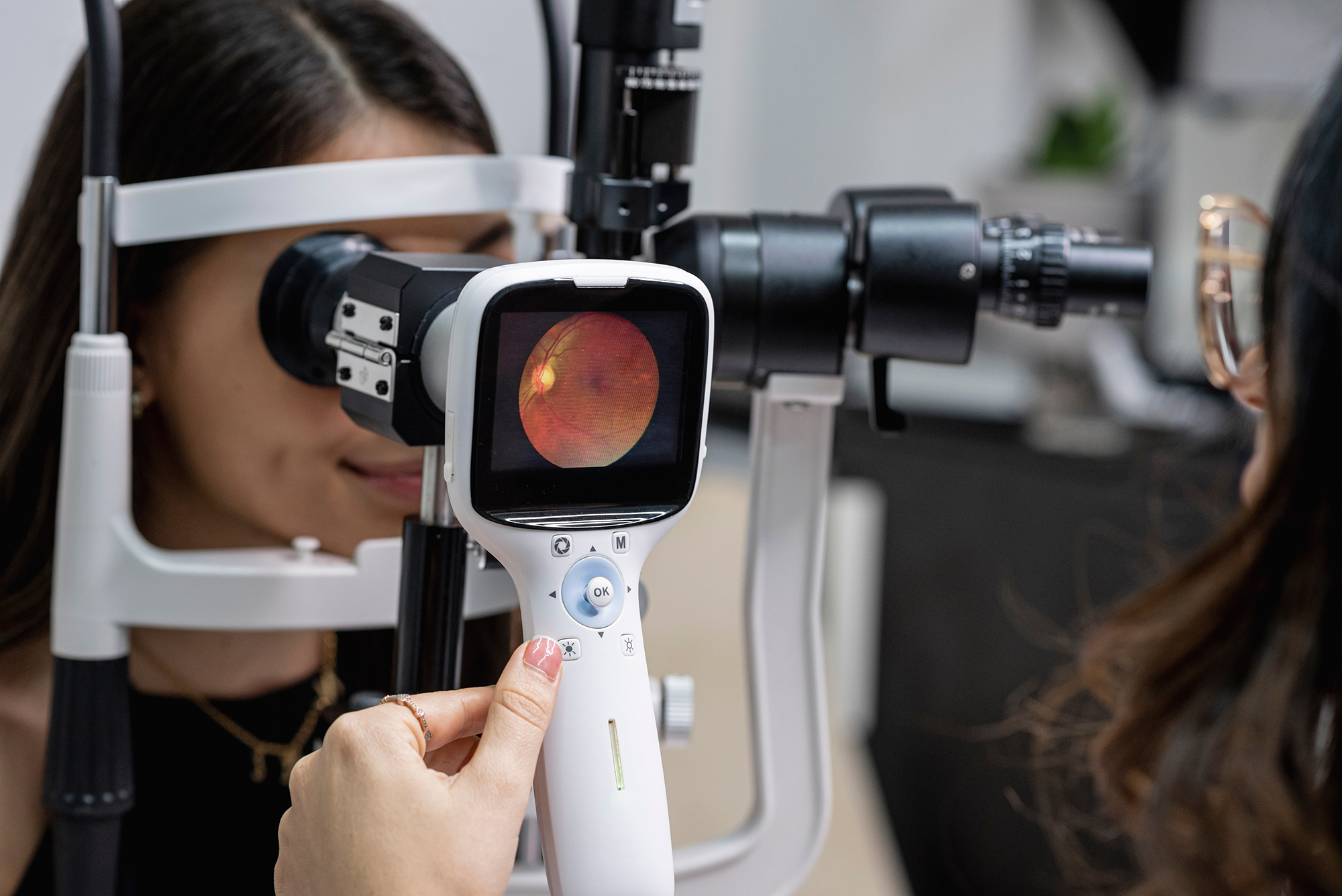
The retina is one of the most important parts of the eye. Located at the back of the eye, this thin layer of tissue is densely packed with light-sensitive cells called photoreceptors.
These cells, called rods and cones, are responsible for taking the information from light and turning it into electrical signals. Once these electrical signals reach the brain via the optic nerve, your brain is able to produce the images you see.
The retina’s structure is complex and includes several layers, each playing a vital role in the vision process. The macula, a small area in the center of the retina, is responsible for your sharp, detailed central vision.
Within the macula is an area called the fovea, which provides the clearest vision and allows you to see details necessary for activities like reading and recognizing faces. Many eye conditions that can lead to vision loss affect this delicate tissue.
By maintaining the health of your retina, you’re preserving one of the most important aspects of your vision and overall quality of life.
Here are some of the most common retinal conditions our specialists diagnose:
Diabetic retinopathy is a complication of diabetes that affects the blood vessels in the retina. Uncontrolled high blood sugar levels can damage these vessels over time, causing them to leak or close off.
When this happens, the retina loses nutrients from proper blood flow. In later stages of the condition, abnormal new blood vessels can grow on the surface of the retina, which can cause more problems with your vision.
Diabetic retinopathy often develops silently, with no early symptoms. As it progresses, you might experience blurry vision, dark or empty areas in your vision, difficulty perceiving colors, and, in severe cases, vision loss.
Regular eye exams are essential for those with diabetes to detect and manage this condition early.
Age-related macular degeneration, sometimes called AMD, is one of the leading causes of vision loss in adults over the age of 50. It affects the macula, the central part of the retina responsible for sharp, detailed vision.
There are two main types of AMD: dry and wet. Dry AMD is the more common form.
This type occurs when the macula thins over time and tiny clumps of protein called drusen build up beneath the retina. This can lead to a gradual loss of central vision.
Wet AMD, though less common, is more severe and can cause rapid vision loss. It occurs when abnormal blood vessels grow under the retina and leak fluid or blood, damaging the macula.
Symptoms of AMD may include blurry or distorted central vision, difficulty reading or recognizing faces, and a need for brighter light when doing close-up work. While there’s no cure for AMD, treatments are available to slow its progression and manage symptoms.
Retinal detachment is a serious and potentially blinding eye condition when the retina separates from the back wall of the eye. There are many reasons why a retinal detachment can happen, such as aging, eye injury, or other eye conditions.
When the retina detaches, it loses its supply of oxygen and nutrients, which can lead to permanent vision loss if not treated promptly. Symptoms of retinal detachment may include:
Retinal vein occlusion (RVO) and retinal artery occlusion (RAO) are conditions that affect the blood vessels supplying the retina. RVO occurs when a vein in the retina becomes blocked, while RAO happens when an artery is obstructed.
Both of these conditions can lead to sudden vision loss in the affected eye. They are often associated with underlying health issues such as high blood pressure, diabetes, or cardiovascular disease.
Symptoms can include sudden, painless vision loss in one eye, which may be temporary or permanent depending on the severity of the blockage and how long it lasts.
At Atlantic Eye Institute, our eye doctors use state-of-the-art diagnostic techniques to evaluate the health of your retina and detect conditions in their earliest stages.
Our comprehensive eye exams include several specialized tests, such as:

Extended ophthalmoscopy is an important part of a complete exam of your retina. To accomplish this, your eye doctor will use specialized lenses and a bright light to examine the entire retina in great detail.
This technique allows your retinal specialist to detect subtle changes in the retina’s appearance, identify areas of concern, and monitor the progression of existing conditions. During this part of an eye exam, your pupils will be dilated so your eye doctor can clearly view the retina.
Your eye doctor will make sure your blood vessels are healthy, check for any changes in the macula, and assess the overall condition of the retina.
Fundus autofluorescence (FAF) involves taking a picture of your retina that provides important information about the health of the retinal pigment epithelium, also called the RPE. The RPE is a layer of cells that is crucial for the healthy functioning of your retina.
This test is non-invasive and uses special light waves to create detailed images of the retina. It shows your eye doctor the areas of damage that may not be visible through other examination methods.
FAF is particularly useful in diagnosing and monitoring conditions like age-related macular degeneration and hereditary retinal diseases. By detecting changes in the retina, FAF helps your doctors identify problems early and track the progression of retinal conditions over time.
B-scan ultrasonography, which is also often simply called B-scan, is a tool eye doctors use when they are unable to get a clear view of the retina. This can be common when patients have dense cataracts or vitreous hemorrhage.
B-scan is an ultrasound technique that provides a two-dimensional, cross-sectional view of the eye and the eye socket, allowing your eye doctor to visualize the retina and other structures even when they can’t be seen directly.
During a B-scan, a small probe is gently placed on the eyelid or surface of the eye, and emits high-frequency sound waves that create detailed images of the interior of the eye. This can be helpful when diagnosing conditions like retinal detachment and tumors or assessing the extent of trauma-related damage.
The first line of defense for macular degeneration is awareness.
A simple test of your vision will alert you to any changes that may indicate a problem with macular degeneration or a worsening of your condition. This common test is known as the Amsler Grid.
One of the first signs of macular degeneration can be wavy, broken or distorted lines OR a blurred or missing area of vision. The Amsler Grid can help you spot these early. Early detection of wet AMD is critical because of laser treatment, when indicated, is most successful when performed before damage occurs. Since dry AMD can lead to the development of wet AMD, most patients should use the Amsler Grid. Check with your eye doctor to find out how often you should use this test. You can download this printable version of the Amsler grid for at-home use.
Wear your reading glasses, if you normally use them and sit about 14 inches away from the screen.
At Atlantic Eye Institute, our retinal specialists offer a wide range of advanced treatments for retinal conditions. Our approach is tailored to each patient’s specific needs, taking into account the nature and severity of their condition.
Here are some of the common treatments we provide:
Retinal injections, also known as intravitreal injections, are essential for the treatment of many retinal conditions, specifically those that involve the abnormal growth of blood vessels or where there is fluid accumulation.
At Atlantic Eye Institute, our retinal specialists offer several types of injectable medications, each of which is designed to address specific retinal issues. Avastin, Eylea, and Eylea HD are anti-VEGF (Vascular Endothelial Growth Factor) drugs that help reduce abnormal blood vessel growth and leakage in conditions like wet AMD and diabetic retinopathy.
Vabysmo is a newer medication that targets two distinct pathways involved in retinal disease, offering potentially longer-lasting effects. Izervay and Syfovre are the latest additions to our options, specifically approved for the treatment of geographic atrophy, an advanced form of dry AMD.
These innovative medications help slow the progression of vision loss in this previously untreatable condition.
For certain inflammatory conditions of the retina and choroid, another layer of the eye, your eye doctor may recommend subtenon injections. This technique involves injecting medication into the space around the eye, beneath the outer layer.
Subtenon injections can deliver anti-inflammatory drugs directly to the specific area while minimizing systemic side effects.
Focal laser treatment is a specific technique used to treat specific areas of the retina in those who have diabetic retinopathy or macular edema. Using a focused laser beam, your retinal specialists can seal leaking blood vessels, reduce swelling, and prevent further damage to the retina.
This targeted approach helps preserve vision while minimizing the impact on surrounding healthy tissue.
For more widespread retinal conditions, such as proliferative diabetic retinopathy, your eye doctor may recommend panretinal photocoagulation. This laser treatment targets a larger area of the outer retina, helping to prevent the growth of abnormal new blood vessels.
While PRP may affect peripheral and night vision somewhat, it’s often crucial in preventing severe vision loss in advanced retinal diseases.
When small retinal tears or holes are detected by your eye doctor, prophylactic barrier laser treatment can save your sight. This procedure creates a controlled scar around the affected area, essentially adhering the retina to the underlying tissue.
By doing so, your eye doctor can prevent the tear from progressing to a full retinal detachment, potentially avoiding the need for more invasive surgeries.